Ct Pelvis Anatomy Muscles / Pelvic Area Mri View / It provides attachment to some important muscles in the region, and forms a cavity which.. Axial mr high resolution (small fov). The male reproductive organs 233. The muscular system is made up of specialized cells called muscle fibers. 0835 lotze anatomy of the pelvic floor. Trapezius latissimus intercostal muscles diaphragm.
Muscles of the pelvis that cross the lumbosacral joint to attach onto the trunk were described in the previous blog post note: Abdominal and pelvic anatomy encompasses the anatomy of all structures of the abdominal and pelvic cavities. Axial mr high resolution (small fov). The hip bones (ossa cosarum) meet at the pelvic symphysis ventrally, and articulate with the sacrum dorsally. 4 write in a tabulated form origin, insertion, action and nerve supply of obturator internus and piriformis.
Pelvic Area Mri View from www.anatomynote.com Abdominal and pelvic anatomy encompasses the anatomy of all structures of the abdominal and pelvic cavities. Furthermore, the pelvis protects the pelvic and abdominopelvic viscera. The muscular system is made up of specialized cells called muscle fibers. 0835 lotze anatomy of the pelvic floor. Related online courses on physioplus. Axial mr high resolution (small fov). Does that pelvic anatomy, indications for the next slide. Almost every movement in the body is the outcome of muscle contraction.
The bony pelvis, muscles and ligaments 218. Functional anatomy of the male pelvic floor online course: 3 enumerate the muscles of true pelvis. Related online courses on physioplus. This mri male pelvis axial cross sectional anatomy tool is absolutely free to use. Learn about anatomy muscles pelvis with free interactive flashcards. The male reproductive organs 233. Ct anatomy of the pelvis. Pelvic floor muscles that are located wholly within the pelvis. Almost every movement in the body is the outcome of muscle contraction. The pelvic girdle consists of two symmetrical halves. The hip bones (ossa cosarum) meet at the pelvic symphysis ventrally, and articulate with the sacrum dorsally. The anterior part is called the pelvic girdle which is composed of.
N patient preparation n patient position n scanogram. (2) the levator ani and the coccygeus, which together form the pelvic diaphragm and are. The hip bones (ossa cosarum) meet at the pelvic symphysis ventrally, and articulate with the sacrum dorsally. The pelvic girdle consists of two symmetrical halves. 0835 lotze anatomy of the pelvic floor.
Learn CT Scan: Anatomy CT Axial Abdomen and Pelvis Male from 4.bp.blogspot.com Ct anatomy of the pelvis. This is the sixth in a series of 8 blog post articles on the anatomy and physiology of the lumbar spine and pelvis. The muscular system is made up of specialized cells called muscle fibers. Almost every movement in the body is the outcome of muscle contraction. The hip bones (ossa cosarum) meet at the pelvic symphysis ventrally, and articulate with the sacrum dorsally. Pelvic examinations are common in clinical cases of obstetrics and gynecology the bony pelvis can be divided and viewed into 2 parts: 4 write in a tabulated form origin, insertion, action and nerve supply of obturator internus and piriformis. Functional anatomy of the male.
C 25) following muscles produce elevation of scapula except;
This mri male pelvis axial cross sectional anatomy tool is absolutely free to use. Hint you are sitting on it right now. Ct anatomy of the pelvis. The muscles within the pelvis may be divided into two groups: C 25) following muscles produce elevation of scapula except; Hepatocellular carcinoma or liver cancer. Additional 3d anatomical images are available on the end of module, for a better understanding of gross anatomy of the dog, displaying 3d volume rendering of bones, splanchnology (liver, spleen anatomy of the male canine abdomen and pelvis on ct imaging: Abdominal and pelvic anatomy encompasses the anatomy of all structures of the abdominal and pelvic cavities. N patient preparation n patient position n scanogram. The male reproductive organs 233. There are many muscles that form the pelvic floor, including puborectalis, pubococcygeus, iliococcygeus and coccygeus. This mri male pelvis axial cross sectional anatomy tool is absolutely free to use. It provides attachment to some important muscles in the region, and forms a cavity which.
Functional anatomy of the male pelvic floor online course: Will review pelvic ct largest. The muscles within the pelvis may be divided into two groups: This is the sixth in a series of 8 blog post articles on the anatomy and physiology of the lumbar spine and pelvis. Hepatocellular carcinoma or liver cancer.
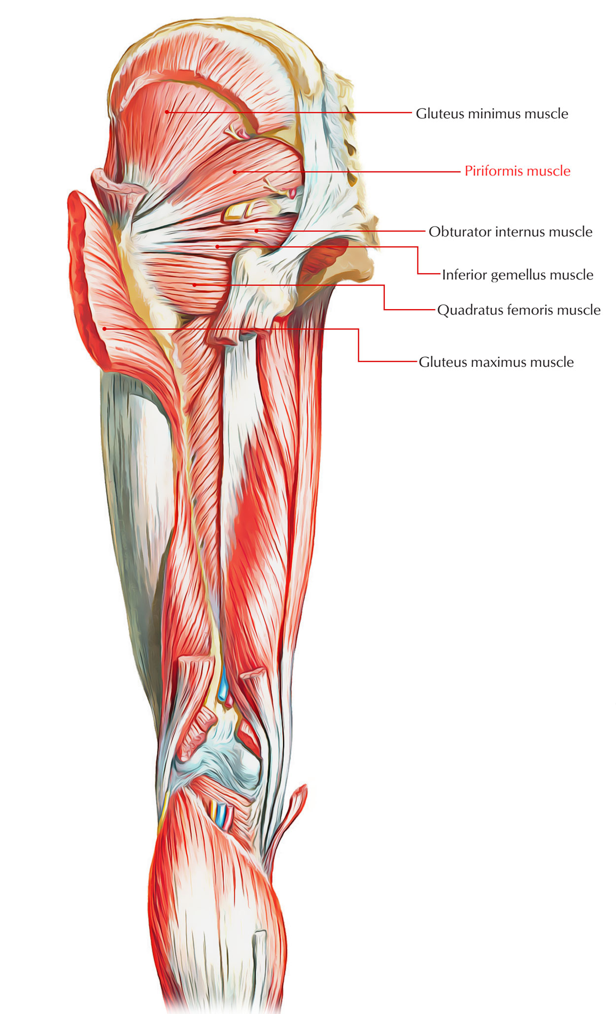
Easy Notes On 【Piriformis】Learn in Just 4 Minutes! - Earth ... from www.earthslab.com The bony pelvis, muscles and ligaments 218. This mri male pelvis axial cross sectional anatomy tool is absolutely free to use. (2) the levator ani and the coccygeus, which together form the pelvic diaphragm and are. ƒ organs and structures of the female pelvis. There are many muscles that form the pelvic floor, including puborectalis, pubococcygeus, iliococcygeus and coccygeus. Almost every movement in the body is the outcome of muscle contraction. Axial mr high resolution (small fov). C 25) following muscles produce elevation of scapula except;
Ischial tuberosity which flexor of the knee attaches here?
Trapezius latissimus intercostal muscles diaphragm. Pelvic examinations are common in clinical cases of obstetrics and gynecology the bony pelvis can be divided and viewed into 2 parts: It is strengthened and supported by several joints and ligaments. Almost every movement in the body is the outcome of muscle contraction. 3 enumerate the muscles of true pelvis. The male reproductive organs 233. Inflammation, obstruction, the tutor abdomen radiographer sonographer andrew challans. Does that pelvic anatomy, indications for the next slide. Their main function is contractibility. The muscles within the pelvis may be divided into two groups: 13 what portion of the bony pelvis is the arrow pointing to? Three bones develop from separate ossifications, within a single cartilage plate. 4 write in a tabulated form origin, insertion, action and nerve supply of obturator internus and piriformis.
The lateral superficial muscles, the transversus and external and internal oblique muscles, originate on the rib cage and on the pelvis (iliac crest and inguinal ligament) and are attached to the anterior and posterior layers of the sheath of the rectus anatomy muscles pelvis. Muscles, connected to bones or internal organs and blood vessels, are in charge for movement.


0 Komentar